

Ever wondered how doctors check if your kidneys are blocked or shrinking without a single X-ray? The answer is renal ultrasound. It is the go-to tool for checking kidney health because it uses sound waves instead of radiation. This makes it safe for pregnant women, children, and anyone who needs frequent check-ups. If you have been referred for this test, you likely have symptoms like back pain, blood in urine, or high blood pressure. Understanding what the scan looks for helps you prepare and interpret your results better.
This guide breaks down exactly how renal ultrasound works, what normal measurements look like, and when doctors might need other tests. We will cover the specific numbers that matter, such as kidney length and resistive index, so you can follow along during your appointment. You will also learn about the limitations of the scan, ensuring you know when a CT scan or MRI might be necessary.
How Renal Ultrasound Works
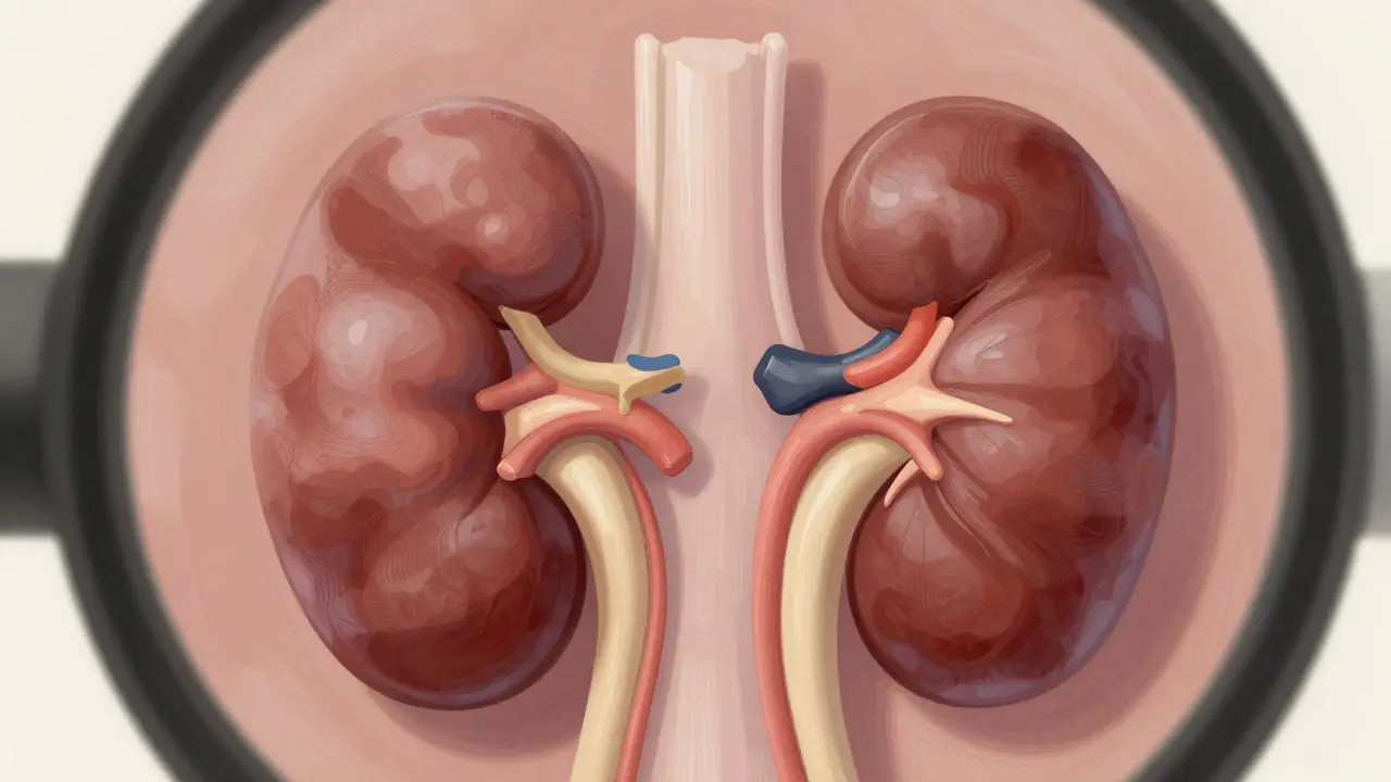
Renal ultrasound is a non-invasive imaging technique that uses high-frequency sound waves to visualize kidney anatomy and detect urinary tract obstructions. Developed in the 1970s, it has become the first-line diagnostic tool worldwide. Unlike CT scans, which use ionizing radiation, ultrasound relies on a transducer placed on your abdomen. This device sends sound waves into your body and bounces them off your organs. The machine then converts these echoes into real-time images on a screen.
The procedure is quick, usually taking only 15 to 30 minutes. You do not need to fast or take special medication beforehand. However, drinking plenty of water before the scan helps fill your bladder, which acts as an acoustic window to see the kidneys more clearly. The main advantage here is safety. Since there is no radiation exposure, doctors can repeat the test as often as needed to monitor conditions like hydronephrosis, which is the dilation of the renal pelvis due to urine accumulation.
Key Measurements and Normal Values
When a radiologist performs the scan, they focus on several specific metrics. These numbers help determine if your kidneys are healthy or under stress. Here is what they are looking for:
- Kidney Length: For adults, the normal range is typically between 9 and 13 centimeters. Significant shrinkage (atrophy) suggests long-term damage, while enlargement might indicate acute blockage or swelling.
- Cortical Thickness: The outer layer of the kidney should be thicker than 1 centimeter. Thinning of this cortex is a sign of chronic kidney disease or prolonged obstruction.
- Renal Pelvis Diameter: A normal anteroposterior diameter is less than 7 millimeters. If this number is higher, it indicates fluid backup, a condition known as hydronephrosis.
- Resistive Index (RI): This is calculated using Doppler ultrasound to measure blood flow. An RI threshold of 0.70 or higher is a strong indicator of obstructive uropathy, with studies showing 86.7% sensitivity and 90% specificity for diagnosis.
| Metric | Normal Value | Abnormal Indication |
|---|---|---|
| Kidney Length | 9 - 13 cm | <9 cm (Atrophy) or >13 cm (Swelling) |
| Cortical Thickness | >1 cm | <1 cm (Chronic Damage) |
| Pelvic Diameter | <7 mm | >7 mm (Hydronephrosis) |
| Resistive Index | <0.70 | ≥0.70 (Obstruction) |
Detecting Obstruction and Stones
One of the primary reasons for a renal ultrasound is to find blockages. Common causes include kidney stones, strictures, or tumors. While ultrasound is excellent at seeing the effects of a blockage, such as swollen kidneys, it is not perfect at seeing the stone itself. Sound waves reflect off dense objects, but small stones can be hidden by bowel gas or bone.
According to the American College of Emergency Physicians, point-of-care renal ultrasound has a sensitivity of about 70% and specificity of 75% when compared to CT scans. This means if you have a large stone causing significant swelling, the ultrasound will likely catch it. However, if the stone is very small (under 3 mm), a CT scan might be needed for confirmation. Despite this limitation, ultrasound remains the preferred initial test because it avoids the approximately 10 mSv of radiation delivered by a standard CT urogram.
Doctors also use Doppler settings to check blood flow. In cases of ureteropelvic junction (UPJ) obstruction, a crossing vessel might be compressing the ureter. Doppler ultrasound can identify these vessels, helping surgeons plan corrective procedures. Additionally, newer techniques like shear-wave elastography measure the stiffness of the kidney tissue. As obstruction pressure increases, kidney tissue becomes stiffer. This provides a quantitative way to assess the severity of the blockage beyond just looking at size.
Ultrasound vs. Other Imaging Modalities
You might wonder why your doctor didn't just order a CT scan immediately. Each imaging method has its strengths. Here is how renal ultrasound stacks up against other common tests:
- CT Urography: This is the gold standard for detecting stones. It can see stones as small as 1-2 mm. However, it involves radiation and often requires contrast dye, which can affect kidney function. It costs significantly more than ultrasound.
- Magnetic Resonance Urography (MRU): MRU offers superior soft-tissue detail and functional information without radiation. But, it is expensive (3-5 times the cost of ultrasound) and takes longer. It is also less sensitive for detecting calcium-based kidney stones.
- Nuclear Renal Scans: These provide detailed functional data about how well each kidney filters blood. They involve radiation and offer less anatomical detail than ultrasound or CT.
Ultrasound wins in specific scenarios. It is the best choice for pregnant women, pediatric patients, and anyone needing serial monitoring. If you are being treated for a UPJ obstruction, your doctor might use weekly bedside ultrasounds to track healing without exposing you to repeated radiation. For obese patients, however, ultrasound can be less effective. Sound waves struggle to penetrate thick layers of fat, which can obscure the view. In these cases, a BMI over 35 may force the medical team to switch to CT or MRI for a clearer picture.

Preparing for Your Appointment
Preparing for a renal ultrasound is straightforward. You do not need to stop taking your regular medications unless told otherwise. The most important step is hydration. Drink two to three glasses of water one hour before your appointment. A full bladder helps push the intestines out of the way, providing a clearer path for the sound waves to reach your kidneys.
During the exam, the sonographer will apply gel to your abdomen and move a handheld probe over the area. You might be asked to lie on your side or back to get different angles. The process is painless, though you might feel slight pressure from the probe. The entire exam usually takes less than 30 minutes. Results are often available immediately, but a radiologist will review the images to provide a formal report.
If you have a history of difficult ultrasounds due to bowel gas, try to avoid carbonated drinks and gas-producing foods like beans or broccoli the day before. While you cannot control all gas production, reducing intake can improve image quality. Remember, the operator's skill matters. Experienced sonographers are better at obtaining clear views and accurate measurements, particularly for the resistive index, which requires precise placement of the Doppler sample gate.
Frequently Asked Questions
Is renal ultrasound painful?
No, the procedure is completely painless. You will only feel the cool sensation of the gel and mild pressure from the probe moving across your skin. There is no needle insertion or radiation involved.
Can ultrasound detect all kidney stones?
Not always. Ultrasound detects about 80% of stones larger than 3 mm. Smaller stones may be missed due to interference from bowel gas or bone. If clinical suspicion remains high despite a negative ultrasound, a CT scan is usually recommended.
What does a high resistive index mean?
A resistive index (RI) of 0.70 or higher suggests increased resistance to blood flow within the kidney. This is often a sign of obstruction or inflammation. It indicates that the kidney is under stress, possibly due to urine backup or vascular issues.
Do I need to fast before the test?
Generally, no fasting is required. In fact, you are encouraged to drink water to fill your bladder. A full bladder improves the visibility of the kidneys by displacing gas-filled intestines.
How often can I have a renal ultrasound?
As often as needed. Because it uses sound waves rather than radiation, there is no cumulative dose limit. Doctors often use it for monthly or even weekly monitoring in patients with known obstructions or post-surgical recovery.
Latest Posts